20.109(F07): Transmission electron microscopy
From Course Wiki

Contents
Introduction
- TEM
- TEM grid
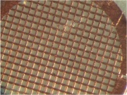 Copper TEM grid with carbon mesh, photographed at 60X magnification
- interp of what's visualized

Protocols
Part 1: TEM sample preparation
- Retrieve your dialyzed sample from the beaker of water and very carefully remove the top clip. The tubing will be slippery and you will be very sad if it slips out of your hand and spills.
- Use your P1000 to sip the sample out of the tubing, squeezing the liquid that's in the tubing toward the tip of the P1000. Place your iridium-coated phage (= nanowires) in an eppendorf tube. You will use only a small aliquot of this solution today but you will need it next time as well so be sure to give the rest of your nanowire sample to the teaching faculty before you leave.
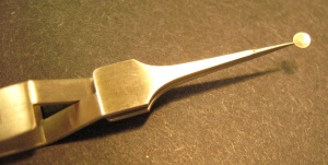
- Place 15 ul of your nanowire solution on the shiny, bright side of the TEM grid that you have balanced in the specialized tweezers.
- Allow the nanowires to settle onto the grid undisturbed for 30'.
- Remove the droplet from the grid with your P200 set to 50 ul.
- Wash the grid by placing adding 15 uL of sterile H2O onto the grid. Immediately remove the water.
- Dry the grid by very gently touching the edge of the grid to a piece of blotting paper in a petri dish. Place the grid (shiny side up) onto the paper to transport to the TEM facility.

Part 2: TEM
DONE!
For next time
- Fig w image
- research proposal paragraph