Difference between revisions of "Protocols for cell culture"
MAXINE JONAS (Talk | contribs) (→Bacteria for aerotaxis studies) |
Steven Nagle (Talk | contribs) (→Bacteria for aerotaxis studies) |
||
| Line 137: | Line 137: | ||
'''Vibrio ordalii:''' | '''Vibrio ordalii:''' | ||
| + | A. From the frozen stock | ||
| + | - Incubation condition: 30C, 200~300 rpm, | ||
| + | - Culture medium: 1/2x 2216 (BD Difco) media (for 1/2x media you can simple dilute 1x with MiliQ water) | ||
| + | - Incubation time: overnight (~20~24 hours) and the cells will be good for the following day | ||
| + | - Culture volume: 2~5 ml in a 15ml culture tube | ||
| + | - Spin the cells at <3,000 rcf to conserve their motility for staining | ||
| + | |||
| + | B. For subsequent culturing from the tube | ||
| + | - Inoculate 20 ul into 2 ml of the culture medium (1/100 dilution) | ||
| + | - Incubate overnight (same as above) | ||
| + | - Do this daily to retrive best motility. | ||
| + | |||
# Grow at 30°C. | # Grow at 30°C. | ||
# From the (overnight) stock mixture: | # From the (overnight) stock mixture: | ||
Revision as of 17:31, 18 March 2014
Preparing Solutions
DMEM reconstitution
For 1 L of DMEM++
- In the hood, combine
- 1 L DMEM
- 100 mL FBS
- 10 mL Penicillin/Streptomycin
- Filter into sterile container.
- Label as DMEM++ and store at 4°C.
3.7% Formaldehyde
- One 16% ampoule
- 3.3 mL 1x PBS
Store in dark at room temperature.
0.1% Triton X-100
- 0.05 g Triton X-100
- 50 mL PBS
Store at room temperature.
1% BSA
- 0.5 g BSA
- 50 mL PBS
Store at 4°C.
10 μM cytochalasin D
- 50 μL 2mM cytoD
- 10 mL PBS
Store at -20°C.
Alexa Fluor dye solution
- 7 μL dye in methanol
- 200 μL PBS
Microsphere cleaning
- For 10 mL of bead solution at a concentration of 5e5 beads/mL: Pipet 1 μL of bead solution and 99 μL of molecular-grade water into centrifuge tube. Vortex.
- Centrifuge the microspheres at 1300 g for 10 minutes to clear the supernatant.
- Remove and discard supernatant. Resuspend the microspheres in 100 μL of water and vortex.
- Centrifuge at 1300 g for 10 minutes.
- Pipet out supernatant and bring into tissue culture hood. Resuspend microspheres in 1 mL of DMEM++ to get concentration of 5e6 beads/mL. (Check in hemocytometer.) Allocate 9 mL of DMEM++ into Falcon tube. Vortex the beads and transfer into the same Falcon tube. Vortex the 10 mL solution. Beads should be at a concentration of 5e5 beads/mL.
- Before pipetting onto cells: vortex the solution, sonicate for 10 minutes to break up aggregates, and warm in hot water bath.
Preparing slides of immobilized microspheres
1. Prepare 1:100 dilutions of fluorescent beads in deionized water:
- In eppendorf tubes, mix 1 mL water with 10 μL beads (stock solution from vendor).
- 3.26 μm red = Spherotech FP-3056-2, Nile Red 1% w/v
- 0.84 μm red = Spherotech FP-0856-2, Nile Red 1% w/v
- PSF red 0.11 μm = Spherotech FP-0256-2, Nile Red 1% w/v
- PSF orange 0.17 μm = Invitrogen P7220, orange (540/560 nm)
2. Prepare microscope slides, so you can act swiftly once agarose is ready and still warm.
3. Prepare 2% agarose gel:
- In glass beaker, mix 50 mL deionized water with 1 g of agarose powder.
- Indeed 1% gel = 1g (agarose) / 100 mL (water)
- Microwave for 60-90 seconds to fully dissolve agarose.
4. Dispense agarose and beads to slide:
- On hot plate (~ 90C to keep agarose as melted as possible), pipet 50 μL agarose gel onto slide (this volume is an extremely rough estimate, given the high viscosity of the agarose gel!)
- With new pipet tip for each slide, then pipet 20 μL fluorescent bead dilution into agarose blurb. Make sure to pipet up and down at least five times, while smearing the agarose and trying to homogenize mix on slide. Avoid bubbles as much as possible.
- Add coverslip, pressing down to homogenize meniscus (which should cover entire coverslip surface, and even 'spill out' a little bit).
5. Let samples dry for two hours. Seal with nail polish or paraffin.
Splitting cells
- Turn on 37°C water bath, open tissue culture hood, turn on light, and spray counter with 70% ethanol. Also wipe (downward) vacuum tube with ethanol. Periodically spray gloves with ethanol throughout the procedure.
- In the hood, to split a single T75 into a single T75, pipette 125 mL of DMEM++ into a separate sterile container. Warm the DMEM++, PBS, and trypsin ontainers in the bath.
- After warming up for 20 minutes, take DMEM++, PBS, and trypsin out of bath. Dry them, wipe with ethanol, and bring into hood.
- Add 25 mL of DMEM++ into the new T75 flask.
- Fetch flask of cells from incubator and make sure they look OK under the inverted microscope. Spray with ethanol and bring into hood.
- Connect a glass Pasteur pipette to the vacuum tube. Use the glass pipette to drain the medium from a corner of the old flask. With the 5 mL pipette, rinse old flask with 4 mL of PBS. Drain with vacuum.
- Pipette 2 mL of trypsin into old flask and close the cap. Set timer for 2 minutes and put in incubator.
- Use the microscope to verify cell detachment.
- Spray flask with ethanol and bring into hood. Pipette 25 mL of DMEM++ into flask of trypsinized cells. Make sure to spread the medium and wash the cells from the walls by pipetting up and down. Using the same pipette, transfer less than 1 mL of cells into an eppendorf tube. Take eppendorf out of hood.
- Transfer ~90 μL of cells and 10 μL of Trypan Blue dye into new eppendorf. Load each chamber of the hemacytometer with ~10 μL of the cell-dye mixture.
- There should be 9 large squares in the field of view. Count the number of cells in the 4 large corner squares and the large center square. Repeat with second chamber and sum the two counts. Multiply by 1000 to estimate the number of cells/mL.
- Calculate the volume required for the desired number of reseeded cells, and pipet that volume into new T75 flask. To plate the MatTek dishes, first add more DMEM++ into the flask of cells such that there are approximately 5x10$ ^4 $ in 0.5 mL. Add 1.5 mL of medium into each MatTek dish. Pipet 0.5 mL of cells into each MatTek. Close the caps, label, and place in incubator.
- Cleanup:
- Vacuum remainder of cells. Drain 50 mL Falcon tube of 50% bleach to wash the system. Disconnect vacuum tubes.
- Put extra DMEM++ and PBS in the fridge. Trysin goes in the -20°C freezer.
- Wipe the counter and close the hood. Clean hemacytometer. Toss eppendorf, old flasks, and gloves in the biowaste container.
Imaging the actin cytoskeleton
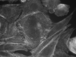
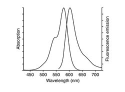
The mushroom toxin phalloidin binds to polymerized filamentous actin (F-actin) much more tightly than to actin monomers (G-actin), and stabilizes actin filaments by preventing their depolymerization. You will take advantage of this strong affinity between phalloidin and F-actin to image the cell's actin cytoskeleton. Phalloidin conjugated with the fluorescent dye Alexa Fluor 568 will be introduced in Chinese hamster ovarian (CHO) cells.

|

|
| Example of a Chinese hamster ovarian cell labeled with Alexa Fluor phalloidin | Excitation and emission spectra for Alexa Fluor 568 |
| |
Wear gloves when you are handling biological samples. |
Procedures for fixing and labeling cells
You are provided with CHO cells, which were prepared as follows: Cells were cultured at 37°C in 5% CO$ _2 $ in standard T75 flasks in a medium referred to as DMEM+++. This consists of Dulbecco's Modified Eagle Medium (DMEM - Invitrogen) supplemented with 10% fetal bovine serum (FBS - Invitrogen), 1% of the antibiotic penicillin-streptomycin (Invitrogen), and 1% of non-essential amino acids (NEAA). The day prior to the fixing/labeling experiments, fibroblasts were plated on 35 mm glass-bottom cell culture dishes (MatTek, equipped with coverslip suited for optical microscopy studies).
Below is the protocol to stain CHO cells with Alexa Fluor 568 phalloidin:
- Start with cells about 60% confluent. This is about the optimum percentage of cell population. If cells are too crowded, they will not stretch properly and show their beautiful actin filaments. Note also that these cells remain alive until the addition of formaldehyde, therefore requiring that any buffer/media added be pre-warmed.
- Pre-warm 3.7% formaldehyde solution, Dulbecco's Modified Eagle's Medium (DMEM) and phosphate buffered saline at pH 7.4 (PBS) in a 37°C water bath. Keep the formaldehyde wrapped in foil to protect from light.
- A key technique to keep in mind when working with live cells - to avoid shocking them with "cold" at 20°C - is to be sure that any solutions you add are pre-warmed to 37°C. We keep a warm-water bath running for this purpose.
- Remove the medium with a pipette and wash the dish 2X with 2 mL of pre-warmed PBS. Pipet up and down into the dish gently to avoid washing away cells.
- Carefully pipet 400 μL of 3.7% formaldehyde solution onto the cells in the central glass region of the dish and incubate for 10 minutes at room temperature. This "fixes" the cells, i.e. cross-links the intracellular proteins and freezes the cell structure.
- Wash the cells 3X with 2 mL PBS (note that this PBS solution no longer needs to be pre-warmed as the cells are dead).
- Extract the dish with 2 mL 0.1% Triton X-100 (a detergent) for 3-5 minutes. (Extraction refers to the partially dissolution of the plasma membrane of the cell.)
- Wash the cells 3X with 2 mL PBS.
- Incubate the fixed cells with 2 mL 1% BSA in PBS for 20 minutes. (BSA blocks the nonspecific binding sites.)
- Wash the cells 2X with PBS.
- Add 200 μL of Alexa Fluor 568 phalloidin solution (pre-mixed in methanol and PBS). Carefully pipet this just onto the center of the dish, cover with aluminum foil, and incubate for 45 minutes at room temperature.
- Wash 3X with PBS.
- You can now store the sample at +4°C (regular refrigerator) in PBS for a few days, wrapped in parafilm and foil. It can also be stored in mounting medium for up to 1 year.
Bacteria for aerotaxis studies
- For them all, we should filter the 2216 media with 0.2-μm filters.
Vibrio alginolyticus:
- Grow at 30°C.
- We should shake it at 300 rpm all week long.
- From the stock mixture:
- Mix 3 mL of 2216 media with 60 μL of stock culture.
- Shake for two hours.
- Then we can monitor the bugs under the microscope.
Vibrio ordalii: A. From the frozen stock - Incubation condition: 30C, 200~300 rpm, - Culture medium: 1/2x 2216 (BD Difco) media (for 1/2x media you can simple dilute 1x with MiliQ water) - Incubation time: overnight (~20~24 hours) and the cells will be good for the following day - Culture volume: 2~5 ml in a 15ml culture tube - Spin the cells at <3,000 rcf to conserve their motility for staining
B. For subsequent culturing from the tube - Inoculate 20 ul into 2 ml of the culture medium (1/100 dilution) - Incubate overnight (same as above) - Do this daily to retrive best motility.
- Grow at 30°C.
- From the (overnight) stock mixture:
- Mix 3 mL of 2216 media with 60 μL of stock culture.
- Shake overnight.
- Then we can monitor the bugs under the microscope.
- These are expected to demonstrate a high aerotactic response.
- Their density is really dense. Too dense for our purposes?
Bacillus subtilis:
- Grow at 37°C.
- Instead of 2216 media, use CAM buffer.
- Their aerotactic response is routinely and convincingly observed in the Stocker lab.
Staining live bacteria
- Obtain two vials of FM 4-64 dye (Invitrogen # T13320) from the center drawer in the 16-352 prep room.
- Obtain one eppendorf of desired bacteria, diluted as-desired.
- Obtain ample fresh warm media for the bacteria, generally at least the same volume as the bacterial sample.
- Obtain 15 mL of HBSS in a new Falcon tube. Sterilize the HBSS as necessary.
- Spin down the two FM 4-64 vials at highest RPM for at least 2 minutes.
- Remove the FM 4-64 vials and spin down the eppendorf of bacteria, and another of water for balance, at highest RPM for 1 minute.
- While the bacteria are spinning, pipette at least 200 uL of HBSS from the Falcon tube into one of the FM4-64 vials (the other was just for balance in the centrifuge) and use the HBSS/FM 4-64 solution to wash and transfer the dye to the Falcon tube.
- Repeat as necessary to extract all dye from one vial.
- Note: This is about 30% greater concentration than specified in the FM product information, but is convenient and works well.
- Remove supernatant from the bacteria and discard.
- Add FM4-64 solution to re-suspend the bacteria pellet. Gently pipette up and down to break up the pellet.
- About 10 times should suffice.
- Incubate at room temperature for 10 minutes.
- Spin down the bacteria at highest RPM for 1 minute.
- Remove supernatant from the bacteria and discard.
- Add fresh warm media to re-suspend the bacteria pellet. Gently pipette up and down to break up the pellet.
- Dispose of waste.
- Return extra FM4-64 concentrate vial to drawer.
- Save FM4-64 dye solution in refrigerator in 20.309 lab.
Optical microscopy lab
Code examples and simulations
- Converting Gaussian fit to Rayleigh resolution
- MATLAB: Estimating resolution from a PSF slide image
- Matlab: Scalebars
- Calculating MSD and Diffusion Coefficients
Background reading
- Geometrical optics and ray tracing
- Physical optics and resolution
- Optical aberrations
- Aperture and field stops
- Optical detectors, noise, and the limit of detection
- Manta G032 camera measurements
- Understanding log plots
