20.109(S22):M1D3
Introduction
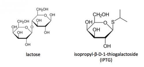
The TDP43 protein is encoded by the TARDBP gene. The entire sequence of the gene is 1242 bases (the protein is 414 amino acids) and composed of four domains: the N-terminal involved in dimerization / oligomerization, two RNA recognition motifs (RRM1 & RRM2) which are required for RNA / DNA binding, and the C-terminal domain involved in protein-protein interactions. In this module, we are interested in ligands that bind the RRM1 and RRM2 domains. For this module, the RRM1 and RRM2 portion of TDP43 was cloned into an expression vector to enable the purification of this segment of the protein. To induce production of TDP43-RRM12 protein from the expression plasmid, a lactose-analogue isopropyl β-D-1-thiogalactopyranoside (IPTG) and arabinose was used to induce expression in BL21(A1) E. coli bacterial cells.
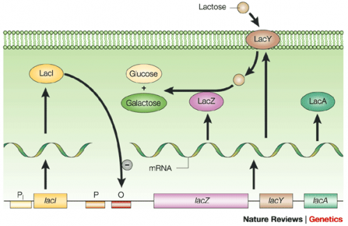
The lac operon is composed of four genes: lacI, lacZ, lacY, and lacA. When lactose is absent, LacI (the protein encoded by lacI) binds to the operator sequence (O) upstream of lacZYA. In the presence of lactose, LacI and lactose form a complex which relieves repression of lacZYA transcription. LacZ is a β-galactosidase that cleaves lactose resulting in glucose and galactose. LacY, a β-galactoside permease, facilitates the transport of lactose across the cell membrane, and LacA, a β-galactoside transacetylase, transfers an acetyl group from acetyl-CoA to β-galactosides.Use of IPTG to induce protein expression is based on the native lac operon used for lactose metabolism in bacterial cells. The native lac operon is a powerful tool in engineering protein expression systems because it enables researchers to control gene expression using inducer molecules. The lacZYA genes are only expressed when lactose is present. If a gene of interest is cloned downstream of the operator sequence, the expression of this gene can be controlled by LacI repression and lactose derepression. To further control the system for protein expression, IPTG is used as a lactose-analog as it is not metabolized by the cells.
Today you will isolate the expressed TDP43-RRM12 protein (remember that the isolated product is actually a truncated version of the TDP43 protein and contains only RRM1 and RRM2!) from the bacterial cells. The TDP43-RRM12 sequence was cloned into an expression vector that contains six histidine codons where the 5' end of the DNA sequence is inserted. Our resultant protein is therefore marked by the presence of these additional encoded residues, also referred to as His-tagged. Histidine has several interesting properties, notably its near-neutral pKa, and His-rich peptides are promiscuous binders, particularly to metals. As an example, histidine side chains help coordinate iron molecules in hemoglobin.
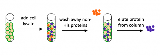
To purify the TDP43-RRM12 proteins present in the bacterial cell, you will use a nickel-agarose resin. The 6xHis-tag on the TDP43-RRM12 protein will bind to the nickel-coated resin, while the other cellular protein will pass through the resin. Remember, the BL21(A1) cells are not only producing the TDP43-RRM12 protein, but also the proteins needed for cellular function and survival. Imidazole is a compound that is also able to bind to nickel and washing the resin with a low concentration solution promotes competition for binding between the imidazole and bound proteins for the nickel-coated resin. Proteins that are non-specifically bound will have a lower affinity for the nickel than imidazole and be washed from the column, whereas the 6xHis-tagged TDP43-RRM12 will remain adhered to the nickel-agarose resin. To elute the TDP43-RRM12 protein from the nickel-coated resin, a high concentration of imidazole is used to out-compete the 6xHis-tag for binding.

Protocols
Part 1: Induce expression of TDP43-RRM12
For timing reasons, the induction steps were completed by the Instructors prior to class.
To ensure the steps included below are clear, please watch the video tutorial linked here: [Bacterial Induction].
- Inoculate 5 mL of LB media containing 50 μg/mL ampicillin with a colony of BL21(A1) cells transformed with pET_MPB_SNAP_TDP43-RRM12.
- Incubate the culture overnight at 37 °C with shaking at 220 rpm.
- Dilute the overnight culture 1:10 in 50 mL of fresh LB media containing 50 μg/mL ampicillin.
- Incubate at 37 °C until the OD600 = 0.6 with shaking at 220 rpm, approximately 4 hours.
- To induce TDP43-RRM12 protein expression, add IPTG to a final concentration of 1 mM and arabinose to a final concentration of 0.2%.
- Incubate 37 °C with shaking at 100 rpm for 2.5 hr.
- To harvest the cells, centrifuge the culture at 3000 g for 15 min at 4 °C.
- Cell pellets were flash frozen in liquid nitrogen, then stored at -80 °C until used for purification.
In your laboratory notebook, complete the following:
- Calculate the volume of ampicillin stock that was added to the LB broth in Step #1. In Step #3.
- Concentration of ampicillin stock = 50 mg/mL.
- Calculate the volume of IPTG stock that was added to the LB broth in Step #5.
- Concentration of IPTG stock = 100 mM.
- Calculate the volume of arabinose stock that was added to the LB broth in Step #5.
- Concentration of arabinose stock = 20%.
Part 2: Purify TDP43-RRM12 protein
Lyse BL21(A1) cells expressing pET_MBP_SNAP_TDP43-RRM12
- Retrieve the BL21(A1) pET_MBP_SNAP_TDP43-RRM12 cell pellet from the -80 °C freezer and leave it on your bench to thaw.
- Add 2ml of cell lysis buffer complete with the following components to each cell pellet. Each cell pellet weighs 0.5 g.
- B-Per bacterial extraction reagent at 4 mL / g of cell pellet
- lysonase at 2 μL / mL of B-Per bacterial extraction reagent
- 1/2 protease inhibitor tablet
- Resuspend the cell pellet in buffer to solubilize the cell pellet and vortex to mix.
- Incubate cell pellet in lysis buffer at room temperature on the nutator at the front desk for 15 minutes.
- Split the lysate evenly across 2 microfuge tubes. It is important that it is split evenly, as the tubes will be used to balance each other within the centrifuge.
- To pellet the cell debris, bring the sample to the front bench so we can centrifuge the lysate at 15,000 g for 30 min at 4 °C.
- Complete the next section (Prepare Ni-NTA affinity column and buffers) during centrifugation.
Prepare Ni-NTA affinity column and buffers
- Obtain a 500 μL aliquot of 50% slurry (Ni-NTA resin) and mix the slurry by inverting the tube several times.
- The slurry is the Ni-NTA column matrix!
- Centrifuge the slurry for 30 sec then remove the supernatent.
- To wash the slurry, add 500 μL of 1X PBS and invert the tube 3 times.
- Cap the bottom of the column and then add the slurry to the column. Cap the top of the column and leave the slurry until ready to use.
- Next, you will prepare buffers for the protein purification protocol: the wash buffer and the elution buffer.
- Wash buffer = 1X PBS containing 10 mM imidazole. You will prepare a total of 10 mL.
- Elution buffer = 1X PBS containing 250 mM imidazole. You will prepare a total of 3 mL.
- Calculate the volume of imidazole stock (concentration = 2.5 M) that should be added to make the wash buffer and elution buffer in the total volumes listed above.
- Confirm your math with the Instructor, then retrieve the 1X PBS aliquots (one for the wash buffer and one for the elution buffer) and an imidazole stock aliquot from the front laboratory bench.
- Prepare your wash buffer and elution buffer by adding the appropriate volume of the imidazole stock to the correct 1X PBS aliquot.
Purify TDP43-RRM12 from cell lysate
- Transfer the supernatent from the centrifuged cell lysate to a fresh microcentrifuge tube.
- Label the microcentrifuge tube containing the cell pellet as "pellet" and give it to the Instructor! This pellet will be used later when protein expression and purity are examined.
- Aliquot 30 μL of the supernatent (from Step #1) to a fresh microcentrifuge tube.
- Label the microcentrifuge tube containing the aliquot as "lysate" and give it to the Instructor! This aliquot will be used later when protein expression and purity are examined.
- Uncap both ends of the column to allow the 1X PBS to run through the column.
- Be sure a 50ml conical tube is placed under the column to collect the waste!
- When the PBS has flowed through, recap the bottom of the column. Do not dry out the resin! The buffer level should be at the same level as the top of the resin.
- Pipet the cell lysate into the prepared Ni-NTA affinity column.
- Be sure that the bottom of the column is capped!
- Attach the cap to the top of the column.
- Incubate the Ni-NTA affinity column on the nutator for 2 hrs at 4 °C.
- Following the incubation, clamp the affinity column into the ring stand.
- Collect the flowthrough from the affinity column.
- Hold a microcentrifuge tube under the column, then remove the bottom cap from the column and collect the liquid that leaves the column.
- Label the microcentrifuge tube as "flowthrough" and give it to the Instructor! This aliquot will be used later when protein expression and purity are examined.
- To wash the Ni-NTA affinity column, add 7.5 mL of 1X PBS containing 10 mM imidazole. Imidazole is available at the bench at 2.5 M
- Hold a microcentrifuge tube under the column, then remove the bottom cap from the column and collect ~250 μL of the liquid that leaves the column.
- Label the microcentrifuge tube as "wash" and give it to the Instructor! This aliquot will be used later when protein expression and purity are examined.
- Allow the rest of the wash to run through. Cap the bottom of the column when the liquid level reaches the top of the resin.
- To elute the TDP43-RRM12 protein from the affinity column, add 1 mL of 1X PBS containing 250 mM imidazole and resuspend by pipetting up and down using a P1000 pipette.
- To collect the purified TDP43-RRM12 protein, hold a 15 mL conical tube under the column, then remove the bottom cap from the column and collect the entire 1 mL of the liquid that leaves the column.
- Repeat Steps #12 and #13 a total of three times to collect 3 mL of eluate.
- Aliquot 30 μL of the eluate (from Step #14) to a fresh microcentrifuge tube.
- Label the microcentrifuge tube as "elution" and give it to the Instructor! This aliquot will be used later when protein expression and purity are examined.
- Lastly, resuspend the slurry from the Ni-NTA affinity column in 250 μL 1X PBS and transfer to a fresh microcentrifuge tube.
- Label the microcentrifuge tube as "slurry" and give it to the Instructor! This aliquot will be used later when protein expression and purity are examined.
In your laboratory notebook, complete the following:
- At several steps in the protein purification procedure, samples are collected that will be used later when protein expression and purity are examined. Consider why each of the samples listed below are saved as controls to measure the success of the purification.
- The pellet from Step #1.
- The lysate from Step #2.
- The flowthrough from Step #9.
- The wash from Step #10.
- The slurry from Step #16.
- What is occurring during the incubation in Step #7?
Part 3: Desalt purified protein
To further prepare the purified protein for the functional assay you will use to assess aggregation, you first need to remove the salt from your protein sample.
- Retrieve a Zeba column from the front laboratory bench.
- Snap off the bottom of a Zeba column, place in a 15 mL conical tube, then loosen the column's cap.
- In the large centrifuge on the instructors bench, centrifuge your columns at 1000 rcf for 2 minutes.
- Transfer the column to a fresh 15 mL conical tube, and then gently add all ~3 mL of your purified protein eluate to the center of the compacted resin.
- Be sure to label the conical tube with your section / team information!
- Centrifuge the column at 1000 rcf for 2 minutes.
- Give the desalted purified protein to the Instructor. Your protein will be stored at 4 °C until the next class session.
Reagents list
- Luria broth (LB) (from BD sciences)
- ampicillin (from Sigma)
- isopropyl β-d-1-thiogalactopyranoside (IPTG) (from Sigma)
- arabinose (from Sigma)
- 2x B-Per bacterial protein extraction reagent (from ThermoFisher)
- lysonase (from Sigma)
- phosphate saline buffer (PBS) (from VWR)
- Ni-NTA agarose (from Qiagen)
- imidazole (from Sigma)
- Zeba column (from Sigma)
- Protease inhibitor tablet (from Roche)
- BL21(A1) bacteria (from Invitrogen)
Next day: Assess purity and concentration of purified TDP43 protein


