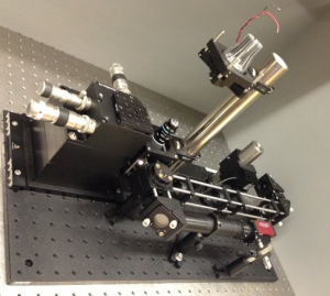
Difference between revisions of "Assignment 1 Overview: Transillumination microscopy"
Juliesutton (Talk | contribs) (→Introduction) |
Juliesutton (Talk | contribs) (→Assignment details) |
||
| (12 intermediate revisions by 3 users not shown) | |||
| Line 6: | Line 6: | ||
| − | |||
| − | |||
| − | |||
| − | |||
| − | |||
| − | |||
| − | |||
| − | |||
| − | |||
| − | |||
| − | |||
| − | |||
| − | |||
| − | |||
==Introduction== | ==Introduction== | ||
[[Image:20.309 130905 InstructorMicroscope1.png|300 px|thumb|right|20.309 microscope|Example 20.309 microscope.]] | [[Image:20.309 130905 InstructorMicroscope1.png|300 px|thumb|right|20.309 microscope|Example 20.309 microscope.]] | ||
| − | Over the next few weeks, you will build an optical microscope using lenses, mirrors, filters, optical mounts, | + | Over the next few weeks, you will build an optical microscope using lenses, mirrors, filters, optical mounts, a CMOS camera, LEDs, and other components in the lab. The work is divided into 5 assignments. Each assignment requires some problem solving, some lab work, some analysis, lots of clear thinking, and an individually written answer sheet turned in on Stellar. All of the items you are expected to turn in are indicated by a pencil symbol in the lab manual. |
{{Template:Assignment Turn In|message =This symbol means that you have to turn something in.}} | {{Template:Assignment Turn In|message =This symbol means that you have to turn something in.}} | ||
| Line 30: | Line 16: | ||
You will work with log-log plots in this assignment and future ones. These seem to confuse everybody. [[Understanding log plots|Read this page]] to remind yourself how log-log plots work. | You will work with log-log plots in this assignment and future ones. These seem to confuse everybody. [[Understanding log plots|Read this page]] to remind yourself how log-log plots work. | ||
| − | Several microscope manufacturers maintain educational websites, including Nikon's [http://www.microscopyu.com MicroscopyU], Olympus' [http://www.olympusmicro.com/primer/index.html Microscopy Primer], and the Zeiss [http://zeiss-campus.magnet.fsu.edu/index.html online microscopy campus]. The content on these sites ranges from basic concepts like [http://www.microscopyu.com/articles/formulas/formulasri.html Snell's law] and [http://www.microscopyu.com/articles/formulas/formulasresolution.html Resolution] to advanced techniques like [https://www.microscopyu.com/tutorials/stochastic-optical-reconstruction-microscopy-storm-imaging | + | Several microscope manufacturers maintain educational websites, including Nikon's [http://www.microscopyu.com MicroscopyU], Olympus' [http://www.olympusmicro.com/primer/index.html Microscopy Primer], and the Zeiss [http://zeiss-campus.magnet.fsu.edu/index.html online microscopy campus]. The content on these sites ranges from basic concepts like [http://www.microscopyu.com/articles/formulas/formulasri.html Snell's law] and [http://www.microscopyu.com/articles/formulas/formulasresolution.html Resolution] to advanced techniques like [https://www.microscopyu.com/tutorials/stochastic-optical-reconstruction-microscopy-storm-imaging super resolution imaging]. |
==Assignment details == | ==Assignment details == | ||
This assignment has 4 parts: | This assignment has 4 parts: | ||
| − | # Part 1: [[Assignment 1, Part 1: Pre-lab questions|Learn about optics and answer a few questions | + | # Part 1: [[Assignment 1, Part 1: Pre-lab questions|Learn about optics and answer a few questions before you start your lab work]]; |
# Part 2: [[Assignment 1, Part 2: Optics bootcamp|Some warm-up lab exercises]]; | # Part 2: [[Assignment 1, Part 2: Optics bootcamp|Some warm-up lab exercises]]; | ||
# Part 3: You will [[Assignment 1, Part 3: Building your transillumination microscope|build a microscope]]; and finally you will | # Part 3: You will [[Assignment 1, Part 3: Building your transillumination microscope|build a microscope]]; and finally you will | ||
| Line 47: | Line 33: | ||
Make sure to include answers to all the following questions: | Make sure to include answers to all the following questions: | ||
| − | Part 1 | + | Part 1: |
| − | # Answers to the pre-lab questions [[Assignment 1, Part 1: Pre-lab questions|listed at the bottom of the Part 1 page]] | + | # Answers to the pre-lab questions [[Assignment 1, Part 1: Pre-lab questions|listed at the bottom of the Part 1 page]]. |
| − | Part 2 | + | Part 2: |
# Turn in your measured focal lengths for each lens A through D. | # Turn in your measured focal lengths for each lens A through D. | ||
| − | # | + | # Make a block diagram of the apparatus (a sketch is fine). You do not need to detail the mechanical construction, but be sure to include any optical elements (light sources, lenses, cameras), as well as the object being measured. Label each component as well as the distances you will be varying (<math>S_i</math> and <math>S_o</math>). Do not label distances that are irrelevant. |
| − | # | + | # List up to three predominant sources of error that affected your measurements of <math> S_o</math> and <math> S_i </math> (i.e. What factors prevented you from making a more accurate or more precise measurement? The answer "human error" does not describe anything specific nor useful, and will not earn you any points.) Estimate the magnitude of each error that you listed. |
| − | # | + | # List one or two predominant sources of error that affected your measurements of <math> h_i </math>. Estimate the magnitude of each error that you listed. |
| − | # | + | # Turn in your table of measured values for <math> S_o, S_i, h_o, h_i, </math> and <math> M </math>. |
| − | + | # Turn in your plot of <math>{1 \over S_i}</math> as a function of <math> {1 \over f} - {1 \over S_o}</math>. | |
| − | # Plot pixel variance vs mean. | + | # Turn in your plot of <math>{h_i \over h_o}</math> as a function of <math>{S_i \over S_o}</math>. |
| − | # | + | # What did you expect each plot to look like based on the theory you learned in class? Did your plots meet your expectations? Why or why not? |
| − | + | # Plot pixel variance ''vs.'' mean. | |
| − | Parts 3 and 4 | + | # Describe how noise varies as a function of light intensity. (Notice that the axes of this plot are in log scale. [[Understanding log plots|Click here]] if you'd like a refresher how to interpret log-log plots.) Did the plot look the way you expected? |
| + | |||
| + | Parts 3 and 4: | ||
| + | |||
# Display an example image of the ruler at each magnification, and | # Display an example image of the ruler at each magnification, and | ||
| − | # Make a table of displaying the nominal magnification | + | # Make a table of displaying the nominal magnification (i.e. the printed number on the objective), the expected magnification (based on the 125 mm tube lens), the object height, the image height, the actual (i.e. measured) magnification and the FOV (see example). Don't forget to include appropriate units. Report the length and width of the FOV (in distance units), not its area (in distance units squared) |
| − | + | ||
# Display an example image of each bead size. | # Display an example image of each bead size. | ||
| − | # | + | # Report the average size and uncertainty of the spheres in each sample, (be sure to include the number of samples measured). |
| − | # In one or two sentences, explain how you chose the number of samples to measure | + | # Discuss how the measured bead sizes compared to the nominal size. |
| − | + | # In one or two sentences, explain how you chose the number of samples to measure. | |
}} | }} | ||
Latest revision as of 17:05, 11 February 2020
Introduction
Over the next few weeks, you will build an optical microscope using lenses, mirrors, filters, optical mounts, a CMOS camera, LEDs, and other components in the lab. The work is divided into 5 assignments. Each assignment requires some problem solving, some lab work, some analysis, lots of clear thinking, and an individually written answer sheet turned in on Stellar. All of the items you are expected to turn in are indicated by a pencil symbol in the lab manual.
| |
This symbol means that you have to turn something in. |
Background reading and resources
You will work with log-log plots in this assignment and future ones. These seem to confuse everybody. Read this page to remind yourself how log-log plots work.
Several microscope manufacturers maintain educational websites, including Nikon's MicroscopyU, Olympus' Microscopy Primer, and the Zeiss online microscopy campus. The content on these sites ranges from basic concepts like Snell's law and Resolution to advanced techniques like super resolution imaging.
Assignment details
This assignment has 4 parts:
- Part 1: Learn about optics and answer a few questions before you start your lab work;
- Part 2: Some warm-up lab exercises;
- Part 3: You will build a microscope; and finally you will
- Part 4: Measure its magnification and the size of some small beads.
You will add fluorescence capability in the next part of the lab.
Submit your work in on Stellar in a single PDF file with the naming convention <Lastname><Firstname>Assignment1.pdf. Here is a checklist of all things you have to turn in:
| |
Make sure to include answers to all the following questions: Part 1:
Part 2:
Parts 3 and 4:
|
- Overview
- Part 1: Pre-lab questions
- Part 2: Optics bootcamp
- Part 3: Build a microscope
- Part 4: Measure stuff
Back to 20.309 Main Page
References

