20.109(S22):M1D6
Part 1: Learn cell culture best practices
One major objective for this experimental module is for you to learn best practices for cell culture using correct sterile techniques. Pay close attention to the demonstration provided by the Instructor!
Review the following resource before you complete the tasks detailed in this exercise: Guidelines for working in the tissue culture room
To ensure the steps included below are clear, please watch the video tutorial linked here: [Cell Culture]. The steps are detailed below so you can follow along!
Preparing tissue culture hood
- The tissue culture hood is partly set up for you. Finish preparing your hood according to the demonstration, first bringing in any remaining supplies you will need, then obtaining the pre-warmed reagents from the water bath, and finally retrieving your cells from the 37 °C incubator.
- Be sure to spray everything (except cells) with 70% ethanol and wipe dry before moving items into the tissue culture hood!
- One of the greatest sources for tissue culture contamination is moving materials in and out of the hood because this disturbs the air flow that maintains a sterile environment inside the hood. Think about what you will need during your experiment to avoid moving your arms in and out of the hood while you are handling your cells.
Collecting cells
- Obtain one ~48 h cultures of MCL-5 cells in T75 flask from the 37 °C incubator.
- Examine your cell cultures after you remove the flask from the incubator.
- Look first at the color and clarity of the media. Fresh media is reddish-orange in color and if the media in your flask is yellow or cloudy, it could mean that the cells are overgrown, contaminated, or starved for CO2.
- Next, look at the cells using the inverted microscope. Note their shape, arrangement, and how densely the cells cover the surface of the flask.
- After you look at your cells, take the flask to your tissue culture hood to begin the splitting procedure.
- With a 10 mL pipet tip, transfer the cell suspension from the T75 flask into a 10 mL conical tube.
- Carefully carry the 10 mL conical tube to the centrifuge and collect the cells at 500 rpm for 3 minutes.
- Return to the tissue culture hood and use the aspirator to remove the media from the cell pellet.
- Attach a fresh pastuer pipet tip to the aspirator tubing before removing the media.
- Be very careful when aspirating the media as it is easy to disturb the pellet!
- With a 5 mL pipet tip, add 5 mL of fresh media to the cell pellet.
- Resuspend the cell pellet by pipetting up-and-down with a 2 mL pipet tip.
- 2 mL pipets are tricky! They fill up quickly. Be careful not to pull up the liquid too quickly or it will go all the way up your pipet into the pipet-aid! If this happens, please alert the teaching faculty rather than returning the pipet-aid to the rack.
- Transfer 90 μL of your cell suspension from the 15 mL conical tube into a labeled eppendorf tube.
Counting cells
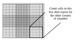
During your work in tissue culture, you will use a hemocytometer to count mammalian cells. More importantly, you will use the cell count information to determine the density of your cultures. A hemocytometer is a modified glass microscope slide that has a chamber engraved with a grid. Stained mammalian cells are loaded into the chamber, which is manufactured such that the area within the gridlines is known and the volume of the chamber is known. These features enable researchers to count the number of cells within a specific volume of liquid.
Using a hemocytometer, you can determine the density (cells per mL) of cell culture.
- Carry the tube with your 90 μL cell suspension aliquot to the center microscope bench and add 10 μL of trypan blue cell stain. Mix by pipetting up and down.
- Carefully pipet 10 μL of the stained cells between the hemocytometer and (weighted) glass cover slip.
- Count the cells that fall within the four corner squares (with a 4 x 4 etched grid pattern), average (i.e. divide by 4), and then multiply by 10,000 to determine the number of cells/mL.
In your laboratory notebook, complete the following:
- Use the information provided to calculate the density of the cell culture.
- Calculate the total number of cells present in the cell suspension.
- Hint: use the density calculated and the total volume for this calculation.
- Calculate the volume of cell suspension that contains 1 M cells.
Part 3: Seed cells for aggragation assay
- Obtain two T75 flasks from the center laboratory bench.
- Clearly label the flasks with the date and your team name. Include the name of the cell line seeded!
- Mix the cell suspension from Part 2.
- Cells settle quickly in conical tubes. It's important you mix before adding cells to the flasks.
- Add 1 M cells into each of the flasks.
- If the cell suspension does not contain 2 M cells, just split the volume of the cell suspension evenly between the two flasks.
- Label the flasks such that the number of cells that were added is easily visible.
- Give any extra cell suspension to the Instructor.
- Add fresh media to each flask such that the final volume equals 12 mL.
- Hint: subtract the volume of cell suspension added from 12 to calculate the volume of fresh media needed.
- Carefully move your flasks to the 37°C incubator
- Clean out the tissue culture hood:
- Aspirate any remaining cell suspensions.
- Dispose of all vessels that held cells in the biohazard waste box and be sure that all sharps are in the sharps jar.
- Remove any equipment or supplies that you transferred into the hood and return to the appropriate location.
- Please leave the equipment that was already there.
- Spray the TC hood surface with 70% ethanol and wipe with paper towels.
- Be sure the paper towels are disposed of in the biohazard waste box!
- Empty the benchtop biohazard bucket into the biohazard waste box.

