Difference between revisions of "20.109(S21):M3D3"
Noreen Lyell (Talk | contribs) (→Part 2: Analyze sequence data) |
Noreen Lyell (Talk | contribs) (→Part 3: Purify IPC protein) |
||
| (40 intermediate revisions by one user not shown) | |||
| Line 5: | Line 5: | ||
==Introduction== | ==Introduction== | ||
| − | + | In the previous laboratory session, you reviewed the mutations that were generated in IPC to create variants. The goal of this was to change calcium binding of IPC by affecting either affinity or cooperativity. Today you will learn how IPC and the IPC variants were expressed and purified, then you will evaluate the success of the protein purification procedure. | |
| − | [[Image:Sp16 M1D4 pRSET vector map.png|thumb|right|350px|''' | + | [[Image:Sp16 M1D4 pRSET vector map.png|thumb|right|350px|'''Schematic of pRSET expression plasmid.''' Modified from Invitrogen manual.]] The genetic sequences that encode the IPC protein and IPC variant proteins are maintained within the pRSET expression vector (recall the cloning exercise from M3D1!). This expression vector contains several features that are important to the expression and purification of IPC and the IPC variants. To enable selection of bacterial cells that carry pRSET_IPC, an antibiotic cassette, specifically an ampicillin marker, is included on the vector. The features most relevant to protein expression and purification are highlighted in the schematic to the right. The T7 promoter drives expression of the gene that encodes IPC (or IPC variant). To ensure that the transcript is translated into a protein, a ribosome binding site (RBS) is included. The ATG sequence serves as the transcriptional start and the 6xHis represents the six-histidine residue tag that is used for protein purification via affinity chromatography. |
| − | + | There are some similarities between the expression system used to purify TDP43-RRM12 in Mod 2 and the system we will use for IPC. First, in both expression systems IPTG is used to induce protein production. As a review, IPTG is a lactose analog that induces expression by binding to the LacI repressor. When bound to IPTG, the LacI repressor is not able to bind to the ''lac'' operator sequence and transcription occurs unimpeded. For more details please review to the [[20.109(S21):M2D1#Introduction |M2D1 Introduction]]! Another similarity is that a 6His tag is used and 6His-tagged IPC and IPC variants will purified using column affinity as shown in the image below. There are also several differences between the expression systems for TDP43-RRM12 and IPC. As you read through the exercises below, consider how these steps are different from those used previously. | |
| − | + | [[Image:Fa20 M2D1 protein purification.png|thumb|550px|center|'''Schematic of affinity separation process.''' For purification, agarose beads (yellow) are coated with nickel (green). When cell lysate is added to the nickel-coated agarose beads, His-tagged protein of interest (blue) adheres to the beads and other proteins in the lysate (orange) are washed from the beads.]] | |
| − | + | ||
| − | + | ||
| − | [[Image: | + | |
| − | + | ||
| − | + | ||
| − | + | ||
| − | + | ||
| − | + | ||
| − | + | ||
==Protocols== | ==Protocols== | ||
| Line 28: | Line 19: | ||
Our communication instructors, Dr. Prerna Bhargava and Dr. Sean Clarke, will join us today for a discussion on preparing a Research proposal presentation. | Our communication instructors, Dr. Prerna Bhargava and Dr. Sean Clarke, will join us today for a discussion on preparing a Research proposal presentation. | ||
| − | ===Part 2: | + | ===Part 2: Prepare protein expression system=== |
| − | + | As mentioned above, IPTG is used to induce protein production in the expression systems for TDP43-RRM12 and IPC; however, the mechanism that drives transcription of the gene that encodes the protein of interest is different. For IPC and the IPC variants, the proteins are expressed using the BL21(DE3)pLysS strain of ''E. coli'', which has the following genotype: F<sup>-</sup>, ''omp''T ''hsd''SB (r<sub>B</sub><sup>-</sup> m<sub>B</sub><sup>-</sup>) ''gal dcm'' (DE3) pLysS (Cam<sup>R</sup>). | |
| + | As mentioned above, pRSET encodes the bacteriophage T7 promoter, which is active only in the presence of T7 RNA polymerase (T7RNAP), an enzyme that therefore must be expressed by the bacterial strain used to make the protein of interest. In BL21(DE3), T7RNAP is associated with a ''lac'' construct. Constitutively expressed ''lac'' repressor (''lac''I gene) blocks expression from the ''lac'' promoter; thus, the polymerase will not be produced except in the presence of repressor-binding lactose or a small-molecule lactose analogue such as IPTG (isopropyl β-D-thiogalactoside). To reduce ‘leaky’ expression of the protein of interest (in our case, IPC), the pLysS version of BL21(DE3) contains T7 lysozyme, which inhibits basal transcription of T7RNAP. This gene is retained by chloramphenicol selection, while the pRSET plasmid itself (and thus IPC) is retained by ampicillin selection. | ||
| + | The pRSET_IPC and pRSET_IPC variants were transformed into chemically competent BL21(DE3)pLysS using heat shock as described previously. To review this method, look back at the information provided on [[20.109(S21):M1D3#Part_3:_Transform_plasmid_from_yeast_into_E._coli |M1D3]]! | ||
| + | [[Image:Sp16 M1D4 protein expression system.png|thumb|center|600px|'''Overview of protein expression system used for IPC purification.''']] | ||
| + | <font color = #4a9152 >'''In your laboratory notebook,'''</font color> complete the following: | ||
| + | *questions about expression system... | ||
| − | ===Part | + | ===Part 3: Purify IPC protein=== |
| − | + | Though the protocol refers only to the purification of IPC, the same procedure was used to purify IPC all of the IPC variants that were assessed in this experiment. | |
| − | + | ||
| − | + | ||
| − | + | ||
| − | + | ||
| − | + | ||
| − | + | ||
| − | + | ||
| − | + | '''Induce expression of IPC''' | |
| − | + | ||
| − | + | #Inoculate 5 mL of LB media containing 50 μg/mL ampicillin and 34 μg/mL chloramphenicol with a colony of BL21(DE3)pLysS cells transformed with pRSET_IPC. | |
| + | #Incubate the culture overnight at 37 °C with shaking at 220 rpm. | ||
| + | #Dilute the overnight culture 1:10 in 50 mL of fresh LB media containing 50 μg/mL ampicillin and 34 μg/mL chloramphenicol. | ||
| + | #Incubate at 37 °C until the OD<sub>600</sub> = ~0.6 with shaking at 220 rpm, approximately 4 hours. | ||
| + | #To induce IPC protein expression, add IPTG to a final concentration of 1 mM. | ||
| + | #Incubate at 25 °C with shaking at 100 rpm overnight. | ||
| + | #To harvest the cells, centrifuge the culture at 3000 g for 15 min at 4 °C. | ||
| + | #Cell pellet was stored at -80 °C until used for purification. | ||
| − | + | <font color = #4a9152 >'''In your laboratory notebook,'''</font color> complete the following: | |
| − | + | *Why is it important that both ampicillin and chloramphenicol are added to the growth media? | |
| − | # | + | |
| − | + | ||
| − | + | ||
| − | + | ||
| − | + | ||
| − | + | ||
| − | + | ||
| − | + | ||
| − | + | ||
| − | + | ||
| − | + | ||
| − | + | ||
| − | + | ||
| − | + | ||
| − | + | ||
| − | + | ||
| − | + | ||
| − | + | '''Lyse BL21(DE3)pLysS cells expressing pRSET_IPC''' | |
| − | + | #Obtain a 2 mL aliquot of room temperature BugBuster buffer and the induced BL21(DE3)pLysS pRSET_IPC cell pellet. | |
| + | #*BugBuster is a bacterial lysis and protein extraction solution, which contains 0.1% bovine serum albumin and 1:200 protease inhibitor cocktail to guard against protein degradation. | ||
| + | #Add 1:1000 of cold nuclease enzyme to the BugBuster buffer. | ||
| + | #Add 600 μL of the BugBuster with nuclease enzyme to the BL21(DE3)pLysS pRSET_IPC cell pellet. | ||
| + | #Resuspend the cell pellet by pipetting until the solution is homogeneous. | ||
| + | #Incubate on the nutator at room temperature for 10 minutes. | ||
| + | #Centrifuge the lysed cell suspension for 10 minutes at maximum speed. | ||
| + | #Transfer the supernatant to a fresh microcentrifuge tube. | ||
| − | + | '''Prepare Ni-NTA affinity column''' | |
| − | + | It is important that all liquid waste generated in the below steps is collected in a designated waste stream due to the presence of nickel in the solution! | |
| − | + | ||
| − | ! | + | |
| − | + | ||
| − | + | ||
| − | + | ||
| − | + | ||
| − | + | ||
| − | + | ||
| − | + | ||
| − | + | ||
| − | + | ||
| − | + | ||
| − | + | #Gently mix the Ni-NTA His-bind resin to fully resuspend, then aliquot 400 μL of the resin into a 15 mL conical tube. | |
| + | #Add 1.6 mL of 1X Ni-NTA Bind Buffer to the Ni-NTA His-bind resin. | ||
| + | #Resuspend the resin by pippeting, then centrifuge at 3300 rpm for 1 minute. | ||
| + | #Carefully remove the supernatant and discard it in the appropriate waste stream. | ||
| − | + | '''Purify TDP43-RRM12 from cell lysate''' | |
| − | + | It is important that all liquid waste generated in the below steps is collected in a designated waste stream due to the presence of imidazole in the solution! | |
| − | + | ||
| − | + | ||
| − | + | ||
| − | + | ||
| − | + | #Add the supernatent from the cell lysis to the prepared Ni-NTA His-bond resin and carefully place on the nutator at at 4°C for 30 minutes. | |
| + | #Centrifuge at 3300 rpm for 1 minute. | ||
| + | #Remove the liquid above the resin and discard it in the appropriate waste stream. | ||
| + | #Add 1 mL of 1X Ni-NTA Wash Buffer to the resin and resuspend. | ||
| + | #Centrifuge at 3300 rpm for 1 minute. | ||
| + | #Remove the liquid above the resin and discard it in the appropriate waste stream. | ||
| + | #Repeat Steps #4-6. | ||
| + | #To collect your purified protein, add 500 μL of 1X Ni-NTA Elute Buffer and resupend. | ||
| + | #Centrifuge at 3300 rpm for 1 minute. | ||
| + | #Transfer the liquid above the resin to a fresh microcentrifuge tube. | ||
| + | #*Please note: your protein is in the liquid at this step! | ||
| + | #Repeat Steps #8-10. | ||
| + | #*Transfer the liquid from the second elution to the same tube used in Step #10. | ||
| + | #*You should have a total of 1 mL of purified protein solution. | ||
| − | + | '''Remove imidazole from purified IPC''' | |
| − | + | ||
| − | + | ||
| − | + | ||
| − | + | ||
| − | + | ||
| − | + | ||
| − | + | ||
| − | + | ||
| − | + | ||
| − | + | ||
| − | + | ||
| − | + | ||
| − | + | ||
| − | + | ||
| − | + | ||
| − | + | ||
| − | + | ||
| − | + | ||
| − | + | ||
| − | + | ||
| − | + | ||
| − | + | ||
| − | + | ||
| − | + | ||
| − | + | ||
| − | + | ||
| − | + | ||
| − | + | ||
| − | + | ||
| − | + | ||
| − | + | ||
| − | + | ||
| − | + | ||
| − | + | ||
| − | + | ||
| − | + | Pilot experiments revealed that imidazole affects the binding curves of inverse pericams. Thus, you will further purify your protein by removing low molecular weight compounds (which includes imidazole!) using a column that removes salt, or desalts, liquid as it passing through a resin. | |
| − | # | + | #Obtain a Zeba column and a 15 mL conical tube. |
| − | # | + | #To prepare the Zeba column, snap off the bottom of a Zeba column and place it into a 15 mL conical tube. |
| − | + | #Centrifuge the column at 2100 rpm for 2 minutes. | |
| − | # | + | #Transfer the column to a fresh 15 mL conical tube, then gently apply your ~1 mL of purified protein solution to the center of the compacted resin. |
| − | + | #Centrifuge the column at 2100 rpm for 2 minutes. | |
| − | # | + | #Transfer the liquid from the 15 mL conical tube to a fresh microcentrifuge tube. |
| − | # | + | #*Please note: your protein is in the liquid at this step! |
| − | + | #From the desalted purified protein solution, aliquot the following amounts: | |
| − | + | #*Add 25 μL to a fresh microcentrifuge tube for examining protein purity using SDS-PAGE. | |
| − | #* | + | #*Add 10 μL to a fresh microcentrifuge tube for examining protein concentration using microBCA. |
| − | + | #Lastly, add a 1:100 dilution of 10% BSA to the remaining desalted purified protein solution. | |
| − | # | + | #*For reference, 10 μL of 10% BSA would be added to 1 mL of protein solution. |
| − | #* | + | |
| − | # | + | |
| − | # | + | |
| − | # | + | |
| − | ===Part | + | ===Part 4: Evaluate purified IPC=== |
| − | + | To evaluate the purified IPC protein, we will use the same methods as when we assessed purified TDP43-RRM12: SDS-PAGE and microBCA (this is a variation of the BCA procedure that is used to measure lower protein concentrations). To review these methods, look back at the information provided on [[20.109(S21):M2D2 |M2D2]]! | |
| − | + | ||
| − | + | ||
| − | + | ||
| − | + | ||
| − | + | ||
| − | + | '''Assess purity using SDS-PAGE''' | |
| − | + | [[Image:Sp21 M3D3 SDSPAGE.png|thumb|500px|center]] | |
| − | + | '''Measure concentration using microBCA''' | |
| − | # | + | #calculate concentration of total protein in each sample... |
| − | + | ||
| − | + | ||
| − | + | ||
| − | + | ||
| − | + | ||
| − | + | ||
| − | + | ||
| − | + | ||
| − | + | #using SDS-PAGE to estimate percentage of total protein that is IPC... | |
| − | + | #might adding different amounts of IPC variants complicate comparisons... how... (move to next day) | |
| − | + | #how could you change experiment such that same amounts of IPC used... (move to next day) | |
| − | + | ||
| − | + | ||
| − | + | ||
| − | + | ||
| − | + | ||
| − | + | ||
| − | + | ||
| − | + | ||
| − | + | ||
| − | + | ||
| − | + | ||
===Part 2: Advance preparation for SDS-PAGE of protein extracts=== | ===Part 2: Advance preparation for SDS-PAGE of protein extracts=== | ||
| Line 244: | Line 166: | ||
</center> | </center> | ||
| − | |||
| − | |||
| − | |||
| − | |||
| − | |||
| − | |||
| − | |||
| − | |||
| − | |||
| − | |||
| − | |||
| − | |||
| − | |||
| − | |||
| − | |||
| − | |||
| − | |||
| − | |||
| − | |||
| − | |||
| − | |||
| − | |||
| − | |||
| − | |||
| − | |||
| − | |||
| − | |||
| − | |||
| − | |||
| − | |||
| − | |||
| − | |||
| − | |||
| − | |||
| − | |||
| − | |||
| − | |||
| − | |||
| − | |||
| − | |||
| − | |||
| − | |||
| − | |||
| − | |||
===Part 4: Protein concentration=== | ===Part 4: Protein concentration=== | ||
| Line 360: | Line 238: | ||
==Reagents list== | ==Reagents list== | ||
| − | + | *Luria-Bertani broth (LB) (from Difco) | |
| − | + | *ampicillin; stock = 100 mg/mL (from Sigma) | |
| − | * | + | *chloramphenicol; stock = 34 mg/mL (from Sigma) |
| − | + | *isopropyl β-d-1-thiogalactopyranoside (IPTG) (from Sigma) | |
| − | + | *BugBuster Protein Extraction Reagent (from EMD Millipore) | |
| − | + | *6X Laemmli sample buffer (from Boston BioProducts) | |
| − | * | + | *4-20% polyacrylamide gels in Tris-HCl (from Bio-Rad) |
| − | + | *TGS buffer: 5 mM Tris, 192 mM glycine, 0.1% (w/v) SDS (pH 8.3) (from Bio-Rad) | |
| − | + | *Precision Plus Dual Color Standard ladder (from Bio-Rad) | |
| − | + | **Molecular weights of ladder bands (linked [http://www.bio-rad.com/en-us/product/prestained-protein-standards?ID=a7b0f9ce-e080-4b51-ab99-4cded66497c1&WT.mc_id=170125006445&WT.srch=1&WT.knsh_id=7417aea6-506f-40cd-96ae-4b6621ba8344&gclid=Cj0KCQjwjer4BRCZARIsABK4QeXhVOAohSuOrR4OKPwHbNnBCWyi5EWgxDPWCbqp67YwI-Qk7AAmMaUaAumyEALw_wcB here]). | |
| − | + | *BioSafe Coomassie G-250 Stain (from Bio-Rad) | |
| − | * | + | *Protein purification supplies (from Novagen/Calbiochem): |
| − | + | ||
| − | * | + | |
| − | + | ||
| − | + | ||
| − | + | ||
| − | + | ||
| − | + | ||
| − | + | ||
| − | *BugBuster Protein Extraction Reagent from EMD Millipore | + | |
| − | + | ||
| − | + | ||
| − | + | ||
| − | + | ||
| − | *6X Laemmli sample buffer from Boston BioProducts | + | |
| − | ** | + | |
| − | + | ||
| − | *Protein purification supplies from Novagen/Calbiochem | + | |
**Ni-NTA His-Bind Resin | **Ni-NTA His-Bind Resin | ||
| − | **1X Ni-NTA Bind Buffer | + | **1X Ni-NTA Bind Buffer; 50 mM NaH<sub>2</sub>PO<sub>4</sub>, pH 8.0; 300 mM NaCl; 10 mM imidazole |
| − | **1X Ni-NTA Wash Buffer | + | **1X Ni-NTA Wash Buffer; 50 mM NaH<sub>2</sub>PO<sub>4</sub>, pH 8.0; 300 mM NaCl; 20 mM imidazole |
| − | **1X Ni-NTA Elute Buffer | + | **1X Ni-NTA Elute Buffer; 50 mM NaH<sub>2</sub>PO<sub>4</sub>, pH 8.0; 300 mM NaCl; 250 mM imidazole |
| − | + | *Zeba Desalt Spin Columns (from Thermo Scientific) | |
| − | *Zeba Desalt Spin Columns from Thermo Scientific | + | *Micro BCA Protein Assay Kit (from Thermo Scientific) |
| − | + | ||
| − | + | ||
| − | *Micro BCA Protein Assay Kit from Thermo Scientific | + | |
| − | + | ||
| − | + | ||
| − | + | ||
| − | + | ||
==Navigation links== | ==Navigation links== | ||
Next day: [[20.109(S21):M3D4 |Evaluate effect of mutations on IPC variants ]] <br> | Next day: [[20.109(S21):M3D4 |Evaluate effect of mutations on IPC variants ]] <br> | ||
Previous day: [[20.109(S21):M3D2 |Examine IPC mutations ]] <br> | Previous day: [[20.109(S21):M3D2 |Examine IPC mutations ]] <br> | ||
Revision as of 17:57, 11 February 2021
Contents
- 1 Introduction
- 2 Protocols
- 3 Reagents list
- 4 Navigation links
Introduction
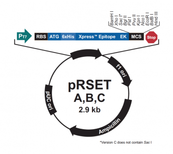
In the previous laboratory session, you reviewed the mutations that were generated in IPC to create variants. The goal of this was to change calcium binding of IPC by affecting either affinity or cooperativity. Today you will learn how IPC and the IPC variants were expressed and purified, then you will evaluate the success of the protein purification procedure.
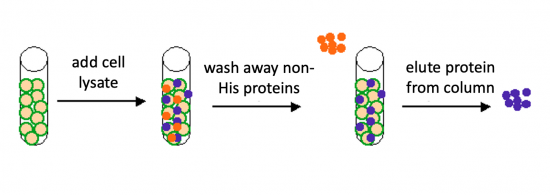
The genetic sequences that encode the IPC protein and IPC variant proteins are maintained within the pRSET expression vector (recall the cloning exercise from M3D1!). This expression vector contains several features that are important to the expression and purification of IPC and the IPC variants. To enable selection of bacterial cells that carry pRSET_IPC, an antibiotic cassette, specifically an ampicillin marker, is included on the vector. The features most relevant to protein expression and purification are highlighted in the schematic to the right. The T7 promoter drives expression of the gene that encodes IPC (or IPC variant). To ensure that the transcript is translated into a protein, a ribosome binding site (RBS) is included. The ATG sequence serves as the transcriptional start and the 6xHis represents the six-histidine residue tag that is used for protein purification via affinity chromatography.There are some similarities between the expression system used to purify TDP43-RRM12 in Mod 2 and the system we will use for IPC. First, in both expression systems IPTG is used to induce protein production. As a review, IPTG is a lactose analog that induces expression by binding to the LacI repressor. When bound to IPTG, the LacI repressor is not able to bind to the lac operator sequence and transcription occurs unimpeded. For more details please review to the M2D1 Introduction! Another similarity is that a 6His tag is used and 6His-tagged IPC and IPC variants will purified using column affinity as shown in the image below. There are also several differences between the expression systems for TDP43-RRM12 and IPC. As you read through the exercises below, consider how these steps are different from those used previously.
Protocols
Part 1: Participate in Comm Lab workshop
Our communication instructors, Dr. Prerna Bhargava and Dr. Sean Clarke, will join us today for a discussion on preparing a Research proposal presentation.
Part 2: Prepare protein expression system
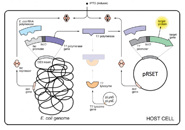
As mentioned above, IPTG is used to induce protein production in the expression systems for TDP43-RRM12 and IPC; however, the mechanism that drives transcription of the gene that encodes the protein of interest is different. For IPC and the IPC variants, the proteins are expressed using the BL21(DE3)pLysS strain of E. coli, which has the following genotype: F-, ompT hsdSB (rB- mB-) gal dcm (DE3) pLysS (CamR).
As mentioned above, pRSET encodes the bacteriophage T7 promoter, which is active only in the presence of T7 RNA polymerase (T7RNAP), an enzyme that therefore must be expressed by the bacterial strain used to make the protein of interest. In BL21(DE3), T7RNAP is associated with a lac construct. Constitutively expressed lac repressor (lacI gene) blocks expression from the lac promoter; thus, the polymerase will not be produced except in the presence of repressor-binding lactose or a small-molecule lactose analogue such as IPTG (isopropyl β-D-thiogalactoside). To reduce ‘leaky’ expression of the protein of interest (in our case, IPC), the pLysS version of BL21(DE3) contains T7 lysozyme, which inhibits basal transcription of T7RNAP. This gene is retained by chloramphenicol selection, while the pRSET plasmid itself (and thus IPC) is retained by ampicillin selection.
The pRSET_IPC and pRSET_IPC variants were transformed into chemically competent BL21(DE3)pLysS using heat shock as described previously. To review this method, look back at the information provided on M1D3!
In your laboratory notebook, complete the following:
- questions about expression system...
Part 3: Purify IPC protein
Though the protocol refers only to the purification of IPC, the same procedure was used to purify IPC all of the IPC variants that were assessed in this experiment.
Induce expression of IPC
- Inoculate 5 mL of LB media containing 50 μg/mL ampicillin and 34 μg/mL chloramphenicol with a colony of BL21(DE3)pLysS cells transformed with pRSET_IPC.
- Incubate the culture overnight at 37 °C with shaking at 220 rpm.
- Dilute the overnight culture 1:10 in 50 mL of fresh LB media containing 50 μg/mL ampicillin and 34 μg/mL chloramphenicol.
- Incubate at 37 °C until the OD600 = ~0.6 with shaking at 220 rpm, approximately 4 hours.
- To induce IPC protein expression, add IPTG to a final concentration of 1 mM.
- Incubate at 25 °C with shaking at 100 rpm overnight.
- To harvest the cells, centrifuge the culture at 3000 g for 15 min at 4 °C.
- Cell pellet was stored at -80 °C until used for purification.
In your laboratory notebook, complete the following:
- Why is it important that both ampicillin and chloramphenicol are added to the growth media?
Lyse BL21(DE3)pLysS cells expressing pRSET_IPC
- Obtain a 2 mL aliquot of room temperature BugBuster buffer and the induced BL21(DE3)pLysS pRSET_IPC cell pellet.
- BugBuster is a bacterial lysis and protein extraction solution, which contains 0.1% bovine serum albumin and 1:200 protease inhibitor cocktail to guard against protein degradation.
- Add 1:1000 of cold nuclease enzyme to the BugBuster buffer.
- Add 600 μL of the BugBuster with nuclease enzyme to the BL21(DE3)pLysS pRSET_IPC cell pellet.
- Resuspend the cell pellet by pipetting until the solution is homogeneous.
- Incubate on the nutator at room temperature for 10 minutes.
- Centrifuge the lysed cell suspension for 10 minutes at maximum speed.
- Transfer the supernatant to a fresh microcentrifuge tube.
Prepare Ni-NTA affinity column
It is important that all liquid waste generated in the below steps is collected in a designated waste stream due to the presence of nickel in the solution!
- Gently mix the Ni-NTA His-bind resin to fully resuspend, then aliquot 400 μL of the resin into a 15 mL conical tube.
- Add 1.6 mL of 1X Ni-NTA Bind Buffer to the Ni-NTA His-bind resin.
- Resuspend the resin by pippeting, then centrifuge at 3300 rpm for 1 minute.
- Carefully remove the supernatant and discard it in the appropriate waste stream.
Purify TDP43-RRM12 from cell lysate
It is important that all liquid waste generated in the below steps is collected in a designated waste stream due to the presence of imidazole in the solution!
- Add the supernatent from the cell lysis to the prepared Ni-NTA His-bond resin and carefully place on the nutator at at 4°C for 30 minutes.
- Centrifuge at 3300 rpm for 1 minute.
- Remove the liquid above the resin and discard it in the appropriate waste stream.
- Add 1 mL of 1X Ni-NTA Wash Buffer to the resin and resuspend.
- Centrifuge at 3300 rpm for 1 minute.
- Remove the liquid above the resin and discard it in the appropriate waste stream.
- Repeat Steps #4-6.
- To collect your purified protein, add 500 μL of 1X Ni-NTA Elute Buffer and resupend.
- Centrifuge at 3300 rpm for 1 minute.
- Transfer the liquid above the resin to a fresh microcentrifuge tube.
- Please note: your protein is in the liquid at this step!
- Repeat Steps #8-10.
- Transfer the liquid from the second elution to the same tube used in Step #10.
- You should have a total of 1 mL of purified protein solution.
Remove imidazole from purified IPC
Pilot experiments revealed that imidazole affects the binding curves of inverse pericams. Thus, you will further purify your protein by removing low molecular weight compounds (which includes imidazole!) using a column that removes salt, or desalts, liquid as it passing through a resin.
- Obtain a Zeba column and a 15 mL conical tube.
- To prepare the Zeba column, snap off the bottom of a Zeba column and place it into a 15 mL conical tube.
- Centrifuge the column at 2100 rpm for 2 minutes.
- Transfer the column to a fresh 15 mL conical tube, then gently apply your ~1 mL of purified protein solution to the center of the compacted resin.
- Centrifuge the column at 2100 rpm for 2 minutes.
- Transfer the liquid from the 15 mL conical tube to a fresh microcentrifuge tube.
- Please note: your protein is in the liquid at this step!
- From the desalted purified protein solution, aliquot the following amounts:
- Add 25 μL to a fresh microcentrifuge tube for examining protein purity using SDS-PAGE.
- Add 10 μL to a fresh microcentrifuge tube for examining protein concentration using microBCA.
- Lastly, add a 1:100 dilution of 10% BSA to the remaining desalted purified protein solution.
- For reference, 10 μL of 10% BSA would be added to 1 mL of protein solution.
Part 4: Evaluate purified IPC
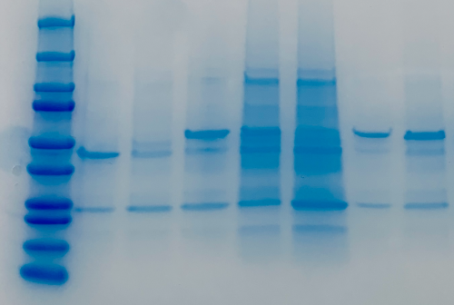
To evaluate the purified IPC protein, we will use the same methods as when we assessed purified TDP43-RRM12: SDS-PAGE and microBCA (this is a variation of the BCA procedure that is used to measure lower protein concentrations). To review these methods, look back at the information provided on M2D2!
Assess purity using SDS-PAGE
Measure concentration using microBCA
- calculate concentration of total protein in each sample...
- using SDS-PAGE to estimate percentage of total protein that is IPC...
- might adding different amounts of IPC variants complicate comparisons... how... (move to next day)
- how could you change experiment such that same amounts of IPC used... (move to next day)
Part 2: Advance preparation for SDS-PAGE of protein extracts
- Last time you measured the amount of cells in each of your samples (-IPTG and +IPTG of the wild-type IPC and one correct mutant). (If you ran cultures overnight, the teaching faculty measured the +IPTG samples for you and posted the results.) Look back at your measurements, and find the sample with the lowest cell concentration. Set aside 15 μL of this sample for PAGE analysis in an eppendorf.
- For your other three samples, you should take the amount of bacterial lysate corresponding to the same number of cells as the lowest concentration sample. For example, if the OD600 of your WT -IPTG sample was 0.05, and the OD600 of your WT +IPTG sample was 0.30, you would take 15 μL of the -IPTG, but only 2.5 μL of the +IPTG sample.
- Next, add enough water so the each sample has 15 μL of liquid in it. You might use the table below to guide your work.
- Finally, add 3 μL of 6X sample buffer to 15 μL of each of your diluted lysates. These will be stored in the freezer until next time.
| Sample Name | OD600 | Sample Volume (μL) | Water Volume (μL) | Total Volume (μL) |
|---|---|---|---|---|
| -IPTG WT | 15 | |||
| +IPTG WT | 15 | |||
| -IPTG mutant | 15 | |||
| +IPTG mutant | 15 |
Part 4: Protein concentration
Part 4A: Prepare diluted albumin (BSA) standards
- Obtain a 0.25 mL aliquot of 2.0 mg/mL albumin standard stock and a conical tube of diH2O from the front bench.
- Prepare your standards according to the table below using dH2O as the diluent:
- Be sure to use 5 mL polystyrene tubes found on the instructors bench when preparing your standards as the volumes are too large for the microcentrifuge tubes.
| Vial |
Volume of diluent (mL) | Volume (mL) and source of BSA (vial) | Final BSA concentration (μg/mL) |
|---|---|---|---|
| A | 2.25 | 0.25 of stock | 200 |
| B | 3.6 | 0.4 of A | 20 |
| C | 2.0 | 2.0 of B | 10 |
| D | 2.0 | 2.0 of C | 5 |
| E | 2.0 | 2.0 of D | 2.5 |
| F | 2.4 | 1.6 of E | 1 |
| G | 2.0 | 2.0 of F | 0.5 |
| H | 4.0 | 0 | Blank |
Part 4B: Prepare Working Reagent (WR) and measuring protein concentration
- Use the following formula to calculate the volume of WR required: (# of standards + # unknowns) * 1.1 = total volume of WR (in mL).
- Prepare the calculated volume of WR by mixing the Micro BCA Reagent MA, Reagent MB, and Reagent MC such that 50% of the total volume is MA, 48% is MB, and 2% is MC.
- For example, if your calculated total volume of WR is 100 mL, then mix 50 mL of MA, 48 mL of MB, and 2 mL of MC.
- Prepare your WR in a 15 mL conical tube.
- Pipet 0.5 mL of each standard prepared in Part 4A into clearly labeled 1.5 mL microcentrifuge tubes.
- Prepare your protein samples by adding 990 μL of dH2O to your 10 μL aliquot of purified protein, for a final volume of 1 mL in clearly labeled 1.5 mL microcentrifuge tubes.
- Add 0.5 mL of the WR to each 0.5 mL aliquot of the standard and to your 0.5 mL protein samples.
- Cap your tubes and incubate at 60°C in the water bath for 1 hour. During this time download the sample data on the Discussion page to practice estimating protein concentration of your samples.
- Following the incubation, the teaching faculty will use the spectrophotometer to measure the protein concentrations of your standards and your purified samples.
- The cuvette filled only with water (H) will be used as a blank in the spectrophotometer.
- The absorbance at 562 nm for each solution will be measured and the results will be posted to today's Discussion page.
- Establish your standard curve by plotting OD562 for each BSA standard (B-H) vs. its concentration in μg/mL.
- Use the standard curve in its linear range (0.5 - 20 μg/mL), and its linear regression in Excel, to determine the protein concentration of each unknown sample (wild-type and mutant IPC).
Reagents list
- Luria-Bertani broth (LB) (from Difco)
- ampicillin; stock = 100 mg/mL (from Sigma)
- chloramphenicol; stock = 34 mg/mL (from Sigma)
- isopropyl β-d-1-thiogalactopyranoside (IPTG) (from Sigma)
- BugBuster Protein Extraction Reagent (from EMD Millipore)
- 6X Laemmli sample buffer (from Boston BioProducts)
- 4-20% polyacrylamide gels in Tris-HCl (from Bio-Rad)
- TGS buffer: 5 mM Tris, 192 mM glycine, 0.1% (w/v) SDS (pH 8.3) (from Bio-Rad)
- Precision Plus Dual Color Standard ladder (from Bio-Rad)
- Molecular weights of ladder bands (linked here).
- BioSafe Coomassie G-250 Stain (from Bio-Rad)
- Protein purification supplies (from Novagen/Calbiochem):
- Ni-NTA His-Bind Resin
- 1X Ni-NTA Bind Buffer; 50 mM NaH2PO4, pH 8.0; 300 mM NaCl; 10 mM imidazole
- 1X Ni-NTA Wash Buffer; 50 mM NaH2PO4, pH 8.0; 300 mM NaCl; 20 mM imidazole
- 1X Ni-NTA Elute Buffer; 50 mM NaH2PO4, pH 8.0; 300 mM NaCl; 250 mM imidazole
- Zeba Desalt Spin Columns (from Thermo Scientific)
- Micro BCA Protein Assay Kit (from Thermo Scientific)
Next day: Evaluate effect of mutations on IPC variants
